Pancreatic Lesions
What is the Pancreas?

The pancreas is an organ located behind the stomach that plays an important double role; it secretes digestive enzymes into the gut, and also regulates blood sugar by the production of insulin and glucagon.
Pancreatic lesions are increasingly being discovered incidentally during scans performed for unrelated reasons. Fortunately, many pancreatic lesions are benign or slow-growing and never cause symptoms.
However, some lesions have the potential to become cancerous over time, making accurate diagnosis and appropriate follow-up important.
What Types of Pancreatic Lesions Occur?
A pancreatic lesion is an abnormal area or growth within the pancreas identified on imaging such as CT, MRI or ultrasound.
Pancreatic lesions broadly fall into two categories: cystic (fluid-filled) lesions and solid lesions.
Pancreatic Cysts
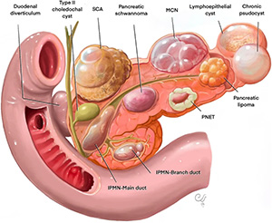
Pancreatic cysts are very common, particularly as people age.
Common types include:
- Simple pancreatic cysts
- Intraductal Papillary Mucinous Neoplasms (IPMN)
- Mucinous Cystic Neoplasms (MCN)
- Serous Cystadenomas
Many pancreatic cysts are completely benign. Others carry a small risk of developing into cancer and may require surveillance or treatment.
Solid Pancreatic Lesions
Solid lesions are less common but often require closer assessment.
Examples include:
- Pancreatic neuroendocrine tumours (NETs)
- Solid pseudopapillary tumours
- Pancreatic adenocarcinoma
- Metastatic tumours
Are Pancreatic Lesions Cancer?
Not necessarily.
Most pancreatic lesions identified incidentally are not cancer.
However, some lesions may represent early cancer or have the potential to become cancerous over time. The challenge is determining which lesions require treatment and which can be safely monitored.
Fortunately, modern imaging and endoscopic techniques allow accurate assessment in most cases.
What Are the Symptoms?
Many pancreatic lesions cause no symptoms and are discovered incidentally.
When symptoms do occur, they may include:
- Upper abdominal pain
- Back pain
- Nausea
- Unexplained weight loss
- Loss of appetite
- New-onset diabetes
- Jaundice (yellowing of the skin or eyes)
Symptoms depend largely on the size, location and type of lesion.
How Are Pancreatic Lesions Diagnosed?
CT Scan and MRI
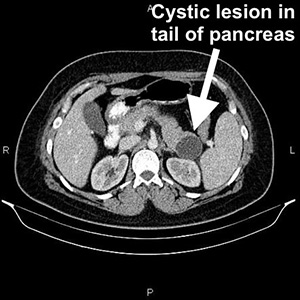
CT and MRI scans are the most important investigations for assessing pancreatic lesions.
These scans help determine:
- The size of the lesion
- Whether it is cystic or solid
- Its relationship to surrounding structures
- Features that may suggest malignancy
Endoscopic Ultrasound (EUS)
Endoscopic ultrasound involves passing a specialised endoscope through the stomach to obtain highly detailed images of the pancreas.
It is often the most accurate test for further characterising pancreatic lesions.
Fine Needle Aspiration (FNA)
During endoscopic ultrasound, a small sample of fluid or tissue can sometimes be obtained for laboratory analysis.
This may help determine the nature of the lesion and whether treatment is required.
How Are Pancreatic Lesions Treated?
Treatment depends on the type of lesion, its size, whether it is causing symptoms and whether there is concern about cancer.
Observation
Many pancreatic cysts can be safely monitored with periodic MRI scans.
The frequency of surveillance depends on the type and size of the lesion.
Surgery
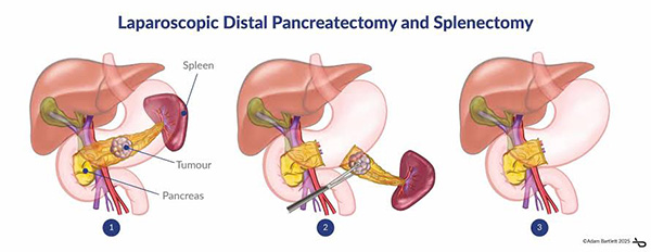
Surgical removal may be recommended when:
- There is concern about cancer
- The lesion is growing
- High-risk features are present
- The lesion is causing symptoms
Depending on the location of the lesion, surgery may involve removal of part of the pancreas or, less commonly, the entire pancreas.
Multidisciplinary Management
Complex pancreatic lesions are often discussed in a multidisciplinary meeting involving surgeons, gastroenterologists, radiologists, pathologists and oncologists to determine the most appropriate management plan.
What Results Can Be Expected?
Most incidentally discovered pancreatic cysts never cause significant problems and can be managed safely with surveillance.
When surgery is required, outcomes are generally excellent for benign and pre-cancerous lesions.
For malignant lesions, outcomes depend on the type of tumour, stage at diagnosis and whether complete surgical removal is possible.
Early detection provides the greatest opportunity for successful treatment.
When Should I Seek Specialist Advice?
You should seek specialist assessment if:
- A pancreatic lesion has been identified on imaging
- You have unexplained abdominal pain
- You have unexplained weight loss
- You develop jaundice
- You have a pancreatic cyst requiring ongoing surveillance
Most pancreatic lesions are not cancerous. However, specialist assessment is important to determine the nature of the lesion and whether treatment or monitoring is required.
Frequently Asked Questions
Are most pancreatic cysts cancerous?
No. Most pancreatic cysts are benign and never become cancerous.
Do all pancreatic lesions require surgery?
No. Many lesions can be safely monitored with periodic imaging.
What is an IPMN?
An Intraductal Papillary Mucinous Neoplasm (IPMN) is a type of pancreatic cyst that produces mucus. Some IPMNs have the potential to become cancerous and therefore require surveillance.
How often do pancreatic cysts need monitoring?
The frequency depends on the size and type of cyst and whether any high-risk features are present.
Can a pancreatic lesion be diagnosed without surgery?
Often yes. Modern CT, MRI and endoscopic ultrasound can frequently determine the nature of a pancreatic lesion without the need for surgery.