Diastasis Recti
What is Diastasis Recti?
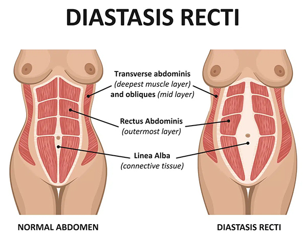
Diastasis recti, also called divarication of the rectus, but commonly referred to as split abdominal muscles or separation of the tummy, is a condition in which the two vertical "six-pack" muscles of the abdomen (rectus abdominis muscles) separate from one another, creating a widening of the connective tissue that normally joins them in the midline.
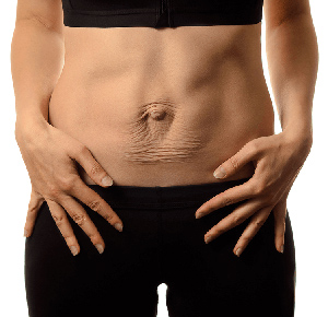
This bulge is particularly noticeable when sitting up, coughing or straining. Unlike a hernia, there is no hole or defect in the abdominal wall. Rather, there is stretching and thinning of the fibrous tissue between the rectus muscles.
Diastasis recti is common after pregnancy but can also occur following significant weight gain, obesity, increasing age, repeated heavy lifting or previous abdominal surgery.
What Causes Diastasis Recti?
The abdominal muscles are normally joined together in the midline by a strong band of connective tissue called the linea alba. When this tissue stretches and weakens, the muscles gradually move apart and the linea alba becomes widened.
Diastasis recti refers to an abnormal widening of the linea alba, resulting in separation of the rectus muscles. While various definitions exist, a separation greater than approximately 2–3 cm is generally considered clinically significant.
Common risk factors include:
- Pregnancy, particularly multiple pregnancies
- Large babies or twin pregnancies
- Significant weight gain or obesity
- Rapid fluctuations in weight
- Chronic coughing
- Heavy lifting and straining
- Previous abdominal surgery
- Age-related weakening of connective tissues
What Are the Symptoms?
Many people with diastasis recti are primarily concerned about the appearance of their abdomen. Others may experience functional symptoms such as:
- A visible bulge or ridge down the centre of the abdomen
- A persistently protruding abdomen despite weight loss
- Core weakness
- Poor posture
- Lower back discomfort or symptoms related to reduced core stability
- Difficulty with exercise or physical activity
- A feeling of instability in the abdominal wall
Symptoms vary considerably, and some people have very few symptoms despite a significant separation.
Is Diastasis Recti a Hernia?
No.
Although the two conditions can look similar, diastasis recti is not a true hernia. In a hernia, there is an actual defect or hole in the abdominal wall through which fat or organs can protrude.
In diastasis recti, the abdominal wall remains intact, but the tissues have stretched and weakened, allowing the muscles to separate.
However, some patients may have both a diastasis recti and a ventral or umbilical hernia at the same time. This is not surprising as both conditions are related to tissue weakness and increased abdominal pressure.
How is Diastasis Recti Diagnosed?
Diagnosis is usually made during a clinical examination.
The separation can often be felt while the patient gently lifts their head from a lying position. A ridge or sausage-shaped bulge in the midline of the upper abdomen is characteristic.
In some cases, ultrasound or CT scanning may be recommended, particularly when there is uncertainty about whether a hernia is also present.
Can Exercise Fix Diastasis Recti?
Targeted physiotherapy and core-strengthening exercises can improve muscle function, strengthen the abdominal wall and reduce symptoms in some patients.
However, while exercise may improve muscle function and reduce symptoms, it is generally unable to reverse a significant separation or restore the connective tissue to its original state once it has stretched.
For patients with mild symptoms, physiotherapy may be all that is required.
When Should Surgery Be Considered?
Surgery may be considered when:
- The separation is severe
- There is significant abdominal wall weakness
- The appearance of the abdomen is distressing
- Symptoms persist despite physiotherapy
- A coexisting ventral or umbilical hernia is present
The decision to proceed with surgery is highly individual and depends on both symptoms and patient goals.
Surgical Treatment
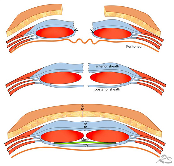
The aim of surgery is to restore the normal anatomy and function of the abdominal wall by bringing the separated rectus muscles back together and reinforcing the weakened midline.
Modern minimally invasive approaches allow reconstruction of the abdominal wall through small incisions, involving restoration of the rectus muscles to the midline and reinforcement of the weakened tissues with mesh.
Click here to read more about surgical treatment options.
When Should I Seek Specialist Advice?
You should consider specialist assessment if you have:
- A persistent bulge down the middle of your abdomen
- Abdominal wall weakness affecting daily activities
- Ongoing symptoms related to poor core stability
- Concerns about the appearance of your abdomen
- A possible associated hernia
A thorough assessment by an experienced surgeon can determine whether you have diastasis recti, a hernia, or both, and help identify the most appropriate treatment options.
Frequently Asked Questions
Can men develop diastasis recti?
Yes. Although commonly associated with pregnancy, diastasis recti also occurs in men, particularly those over the age of 50, following significant weight gain, obesity or long-standing increases in abdominal pressure.
Will diastasis recti get worse over time?
In some patients the separation remains stable, while in others it may gradually widen over time, particularly if underlying risk factors persist.
Can diastasis recti cause a hernia?
Diastasis recti itself is not a hernia, but the two conditions frequently coexist because both are associated with weakness of the abdominal wall tissues.